Radiology Services
- Home
- Radiology Services
Get accurate and trusted diagnostic imaging services at Unihealth Southwoods Hospital in Southwoods, Biñan, Laguna. We serve patients from Biñan, Sta. Rosa, San Pedro, Cabuyao, and surrounding areas in Laguna.
Our Radiology Department offers a comprehensive range of diagnostic imaging services, from standard Chest PA X-ray, high-resolution Whole Abdomen Ultrasound, precise CT-Scan with and without contrast, and advanced MRI scan with and without contrast. Whether you’re looking for an X-ray, ultrasound, or MRI scan
in Biñan Laguna, our department is equipped to handle your diagnostic needs.
Discover our complete list of imaging services below.
Quick guide
Table Of Contents
Services Offered
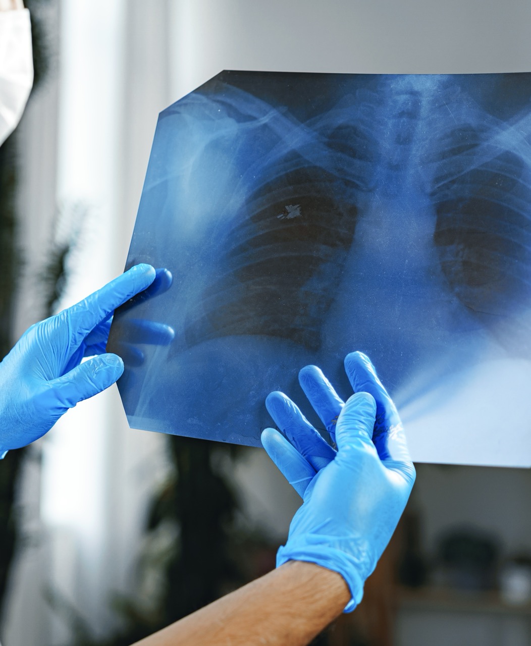
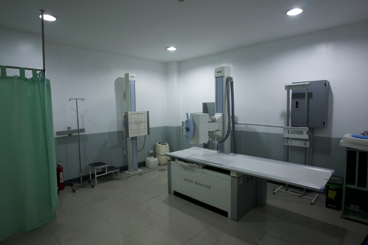
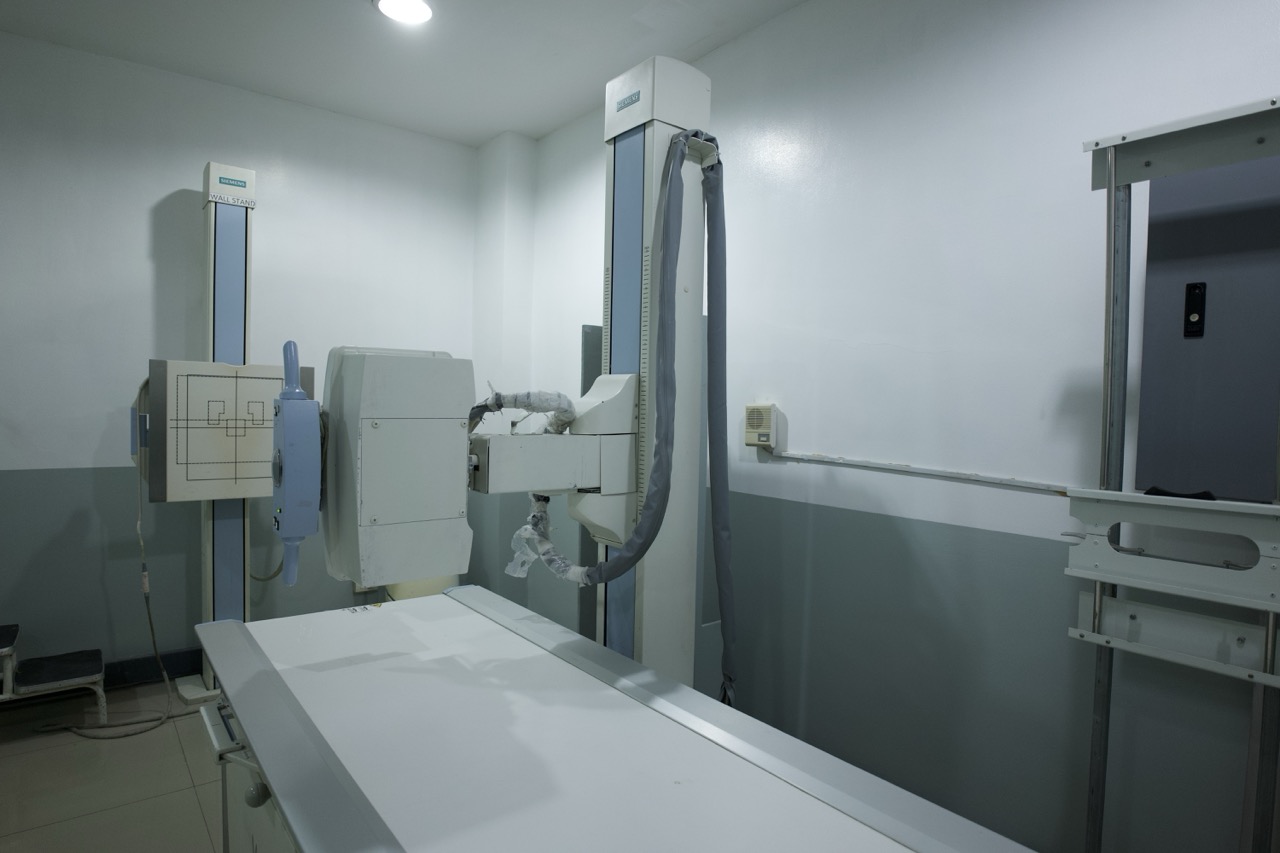
X-ray
X-ray is a non-invasive imaging technique that uses low dose radiation to be able to view bones for possible fractures, dislocations, or deformities, view the chest to see the lungs and heart to check their size and condition, and to check for foreign objects.
Our Digital X-ray machine emits up to 90% lower radiation than conventional X-ray machines, making it a safer option for patients. If you’re looking for Chest X-ray
services in the Philippines at a hospital with modern, low-dose equipment, our Radiology Department in Biñan, Laguna is here to help.
Available X-ray Services:
See List
Abdomen
Ankle
Apico
Babygram
Arm APL
Chest
Cervical
Elbow
Femur
Foot
Hand
Knee
Kub Adult
Kub Pedia
Lumbosacral
Nasal Bone
Paranasal Sinuses
Pelvis
Forearm
Shoulder
Scapula Y
Sunrise/merchant
Skull AP Lateral
Scoliotic Series
Thoracic AP/lateral
Thoracolumbar Spine
Thoracic Cage
Tibia/fibula Leg
Thigh APL
Wrist APL
Whole Spine APL
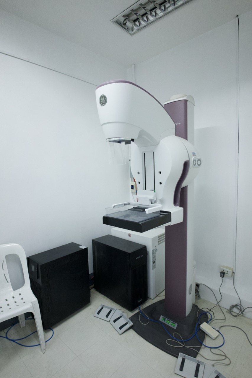
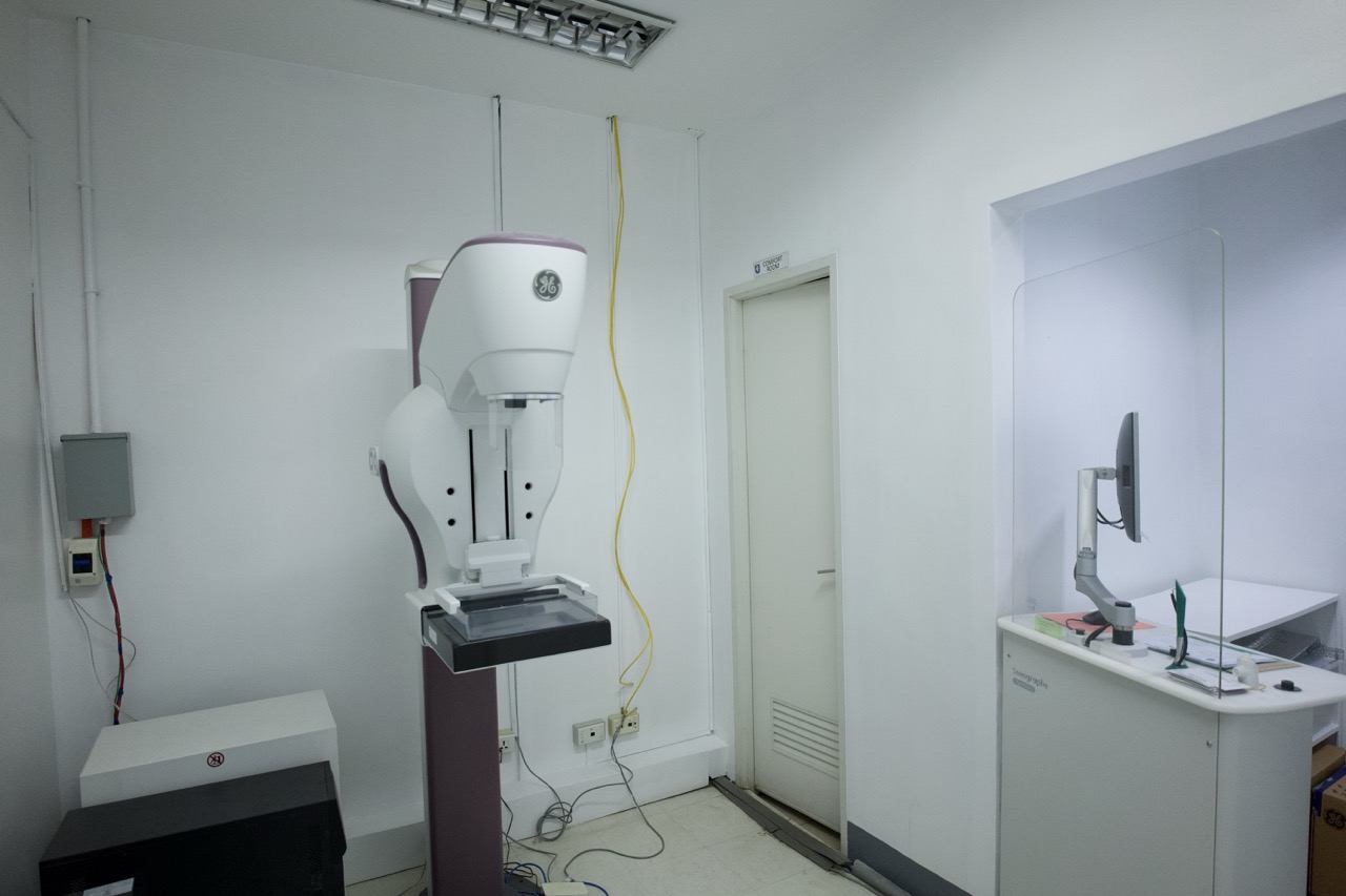
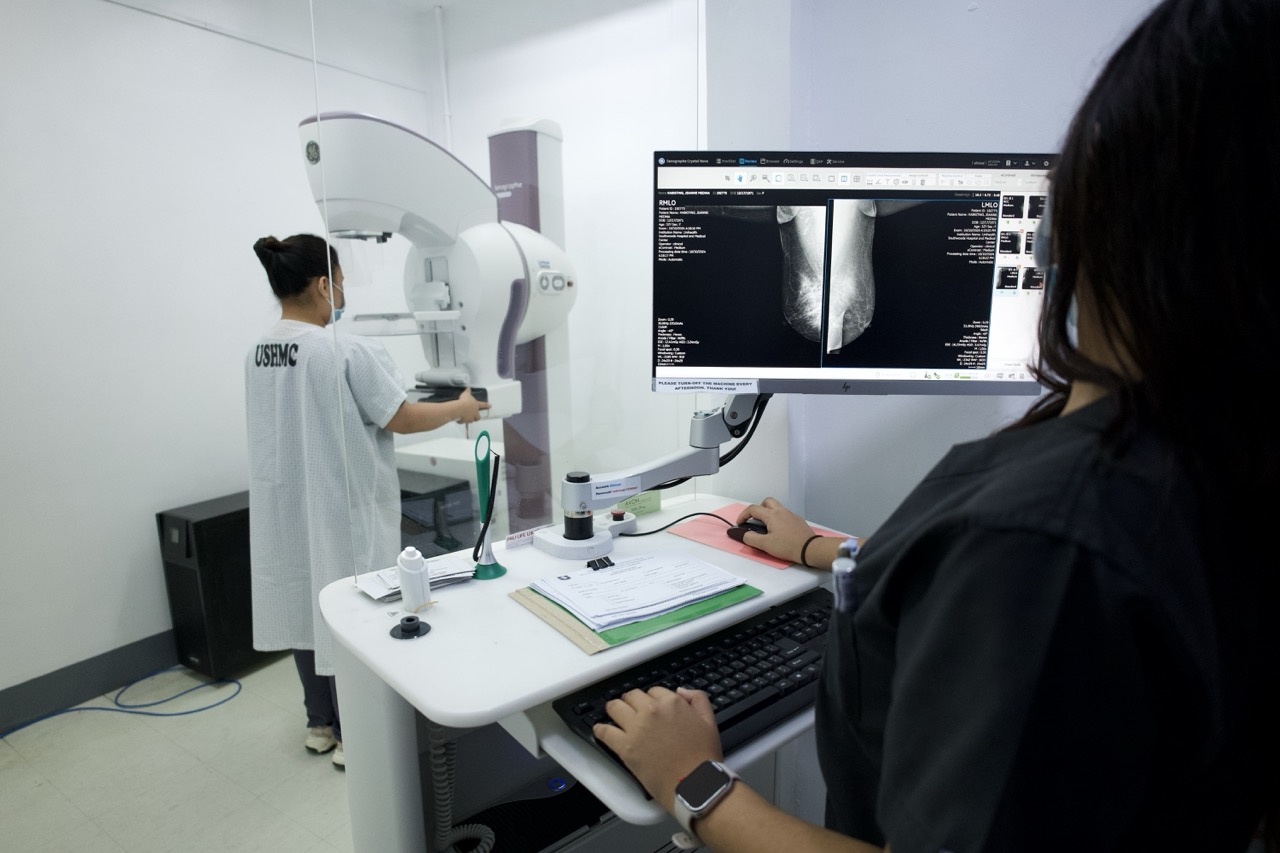
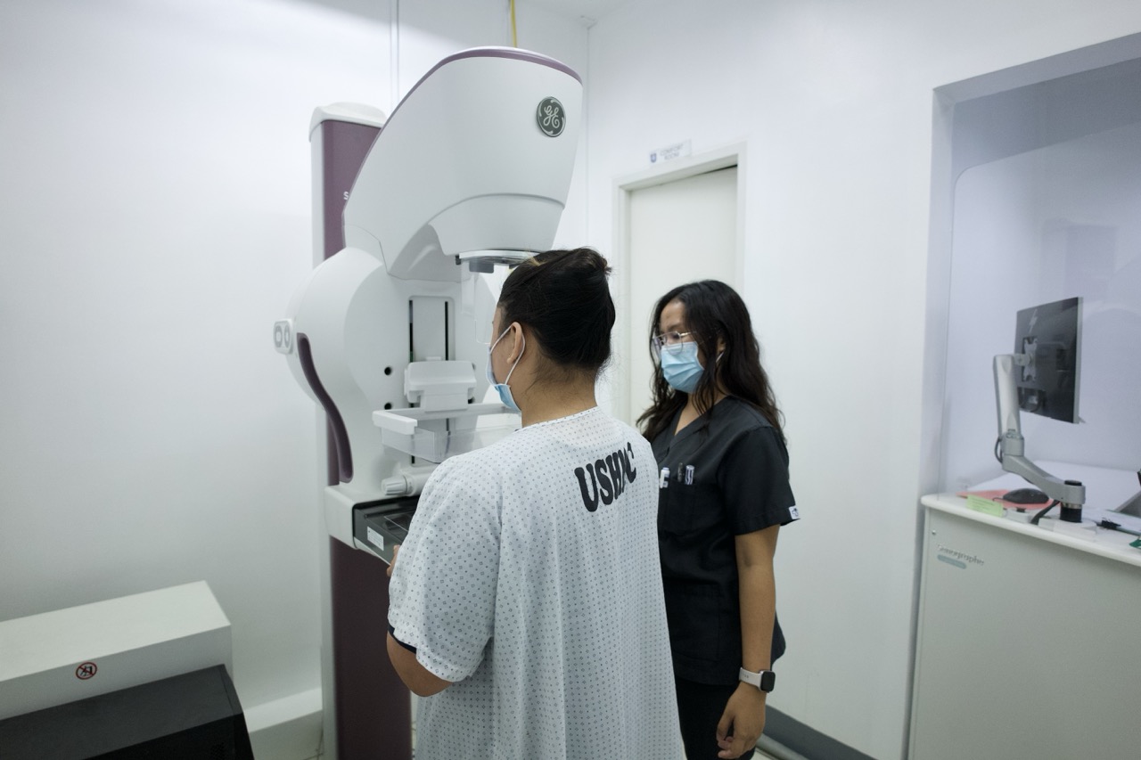
Mammogram
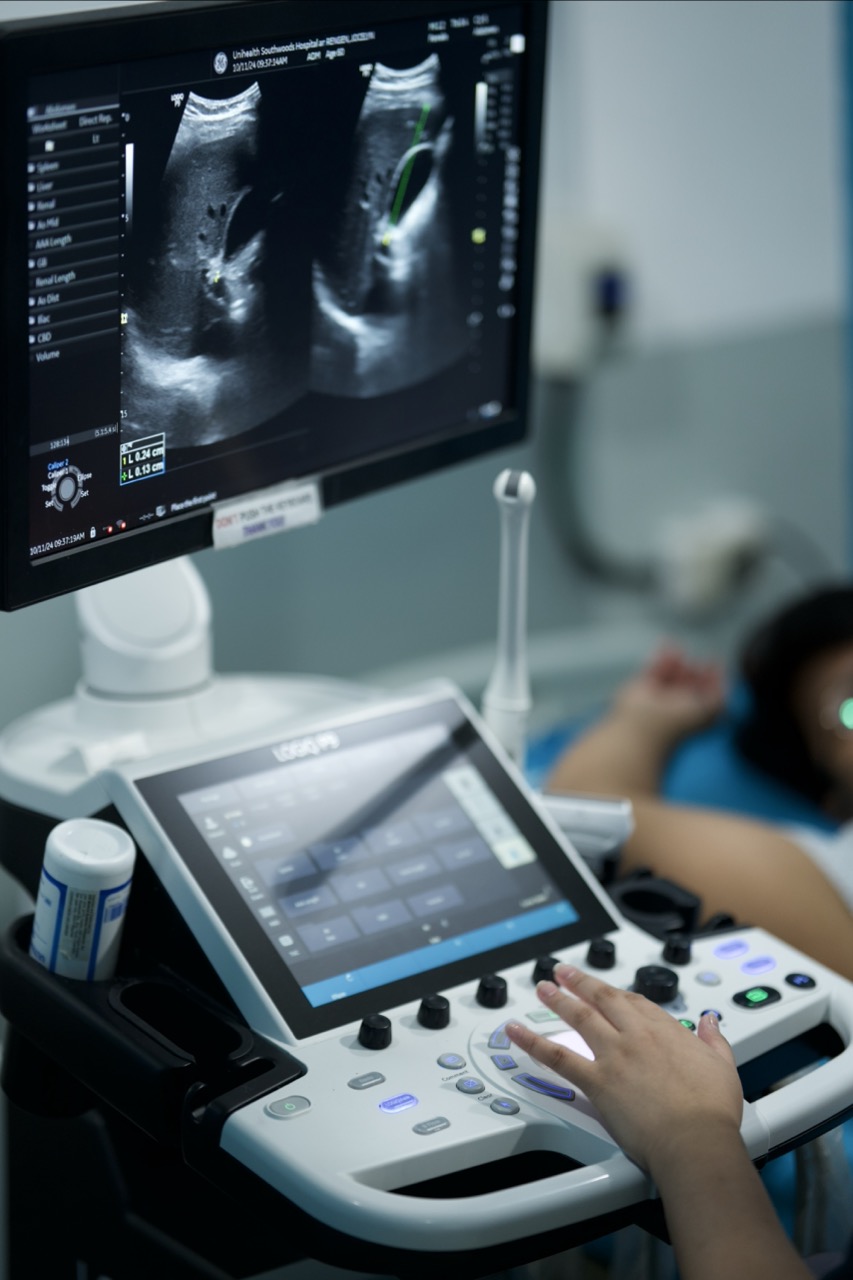
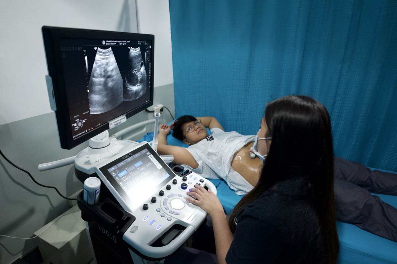
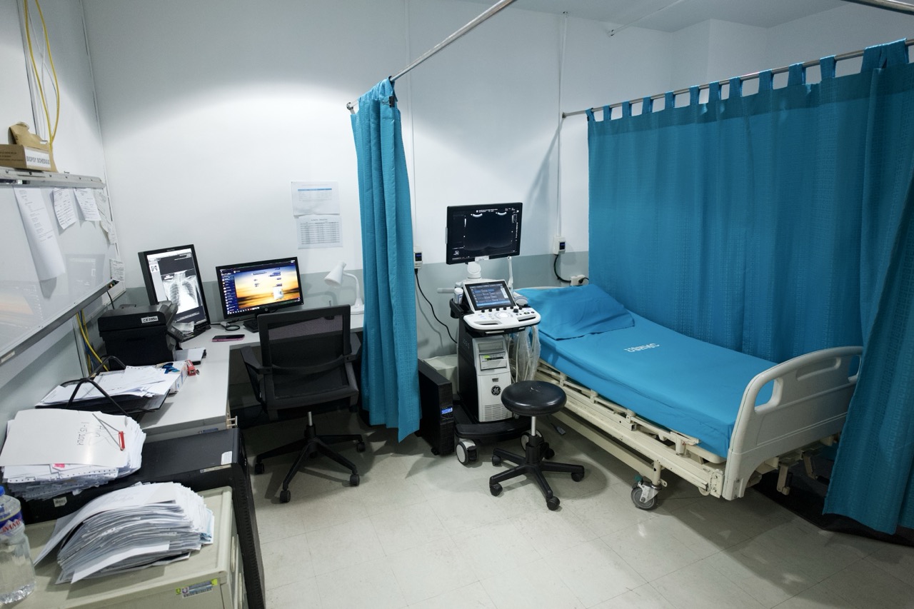
Ultrasound Imaging
Ultrasound, also called Sonographya involves in exposing part of the body to high frequency soundwaves to produce pictures of the inside of the body.
- Ultrasound Examination - do not use ionizing radiation (as used in X-Rays).
- Because ultrasound images are captured in real-time, they can show the structure and movement of the body’s internal organs, as well as blood flowing through blood vessels.
We offer a wide range of ultrasound services at our facility, including Whole Abdomen Ultrasound, Breast Ultrasound, and Thyroid Ultrasound. Our ultrasound clinic serves patients from Biñan, Sta. Rosa, San Pedro, and throughout Laguna.
Available Ultrasound Services:
See List
Appendix
Axillary
Breast
Chest Bilateral
Chest Mapping (unlatateral)
Cranial
Gallbladder
HBT
HBT-Pancreas
Inguinal Area
Kidney
Kub
Kub-Prostate
LGBP
Liver
Neck (B)
Thyroid
Pancreas
Parotid
Prostate
Scrotal Doppler
Scrotum Testis
Inguino Scrotal
Spleen
Umbilical
Upper Abdomen
Lower Abdomen
Urinary Bladder
Whole Abdomen
MSK
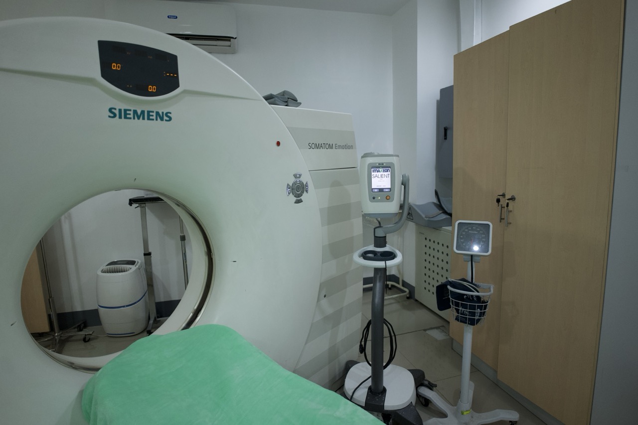
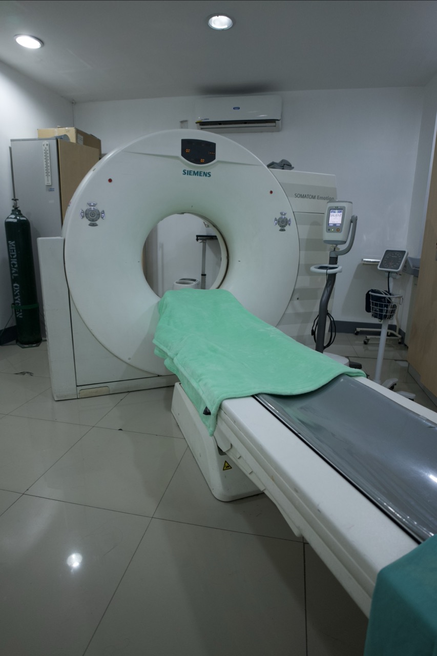
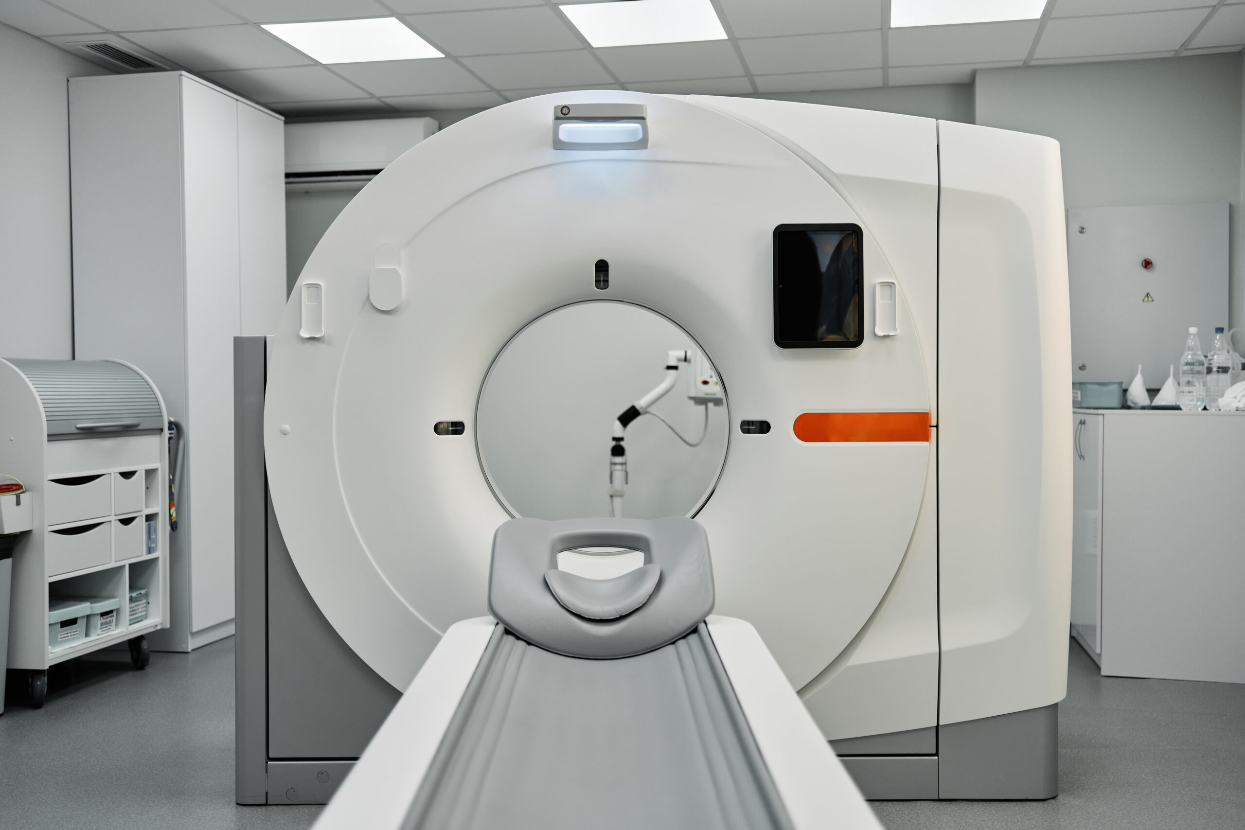
CT-Scan Plain
A Computed Tomography (CT) scan is actually an advanced X-ray scan that makes detailed, cross-sectional slices of internal structures of the body. It is usually used to view hard or more dense structures in the body such as checking for any bone trauma or acute internal injuries without using contrast dye.
- CT Imaging - During CT-Scan, an X-ray tube rotates around the patient, generating multiple slices that are reconstructed into images by the computer. Various scanning protocols are used for different body parts and clinical indications.
Available CT-Scan Plain Services:
See List
Cranial Plain
Chest Plain
Whole Abdomen Plain
Paranasal Plain
Cervical Plain
Lumbo-sacral Plain
Thoracic Spine Plain
Orbit Plain
Maxillo Facial Plain
Ankle Plain
Elbow Plain
Foot Plain
Knee Plain
Shoulder Plain
Leg Plain
Temporal Plain
Forearm Plain
Hip Plain
Arm Plain
Mandible Plain
Stonogram
CT-Scan with Contrast
A Computed Tomography (CT) scan with Contrast uses an iodine-based contrast dye which is injected into the bloodstream to highlight and help visualize organs, tissues, blood vessels and blood flow, allowing for clearer and more detailed images. It also shows infections, tumors, and whether organs are functioning properly. It helps identify and differentiate tumors, abscesses, or blockages from normal looking soft tissue on a plain scan.
Available CT-Scan with Contrast Services:
See List
Cranial With Contrast
Chest With Contrast Estimate
Whole Abdomen With Contrast
Whole Abdomen Triple Contrast Estimate
Thoracic With Contrast
Cervical With Contrast
Whole Abdomen Triphasic
Pulmo Angiogram
Brain Angiogram
Urogram
Aortogram
We understand that CT-Scan prices are an important consideration for patients. For inquiries about our CT-Scan with Contrast price in Laguna, please call our Information Department at +63 925 310 5666.
MRI Plain
A Magnetic Resonance Imaging (MRI) scan is an advanced scan that creates the highest resolution images of soft tissues using very strong magnets along with radio waves. It does not use any radiation and it is usually used to check for tumors, nerve compression, ligament tears, brain injury, strokes, and the functionality of different internal organs such as the brain, liver, and uterus.
Available MRI Plain Services:
See List
Extremities Plain
Cervical Plain
Lumbar Plain
Thoracic Plain
Cranial Plain
Pituitary Plain
Pelvis Plain
Whole Abdomen Plain
Brain w/ MRA (Circle of Willis) Plain
Brain w/ Pituitary Plain
MRCP Plain
MRA Plain (Circle of Willis)
Orbits Plain
Upper/lower Abdomen Plain
Whole Abdomen Plain
Wab w/ Mrcp Plain
Whole Spine Plain

MRI with Contrast
A Magnetic Resonance Imaging (MRI) scan with Contrast uses a Gadolinium-based contrast agent. Gadolinium has magnetic properties that help provide comprehensive, enhanced visualizations of blood flow, inflammation, and abnormal growths such as tumors or lesions. It reveals areas of inflammation or active disease more clearly than a plain MRI.
Available MRI with Contrast Services:
See List
Extremities With Contrast
Brain w/ Contrast
Pelvis w/ Contrast
Pituitary w/ Contrast
Brain w/ MRA w/ Contrast (Magnetic Resonance Angiogram)
Cervical w/ Contrast
Lumbar Spine w/ Contrast
Thoracic Spine w/ Contrast
MRCP w/ Contrast
Orbit w/ Contrast
WAB w/ Contrast With MRCP
WAB w/ Contrast
Brain w/ Contrast and Orbit w/ Contrast
We offer a range of MRI scans with contrast, including Cranial MRI or Brain MRI with and without contrast. For details on our MRI scan price in the Philippines or specific scans like Cranial MRI price, please contact our Information Department at +63 925 310 5666.
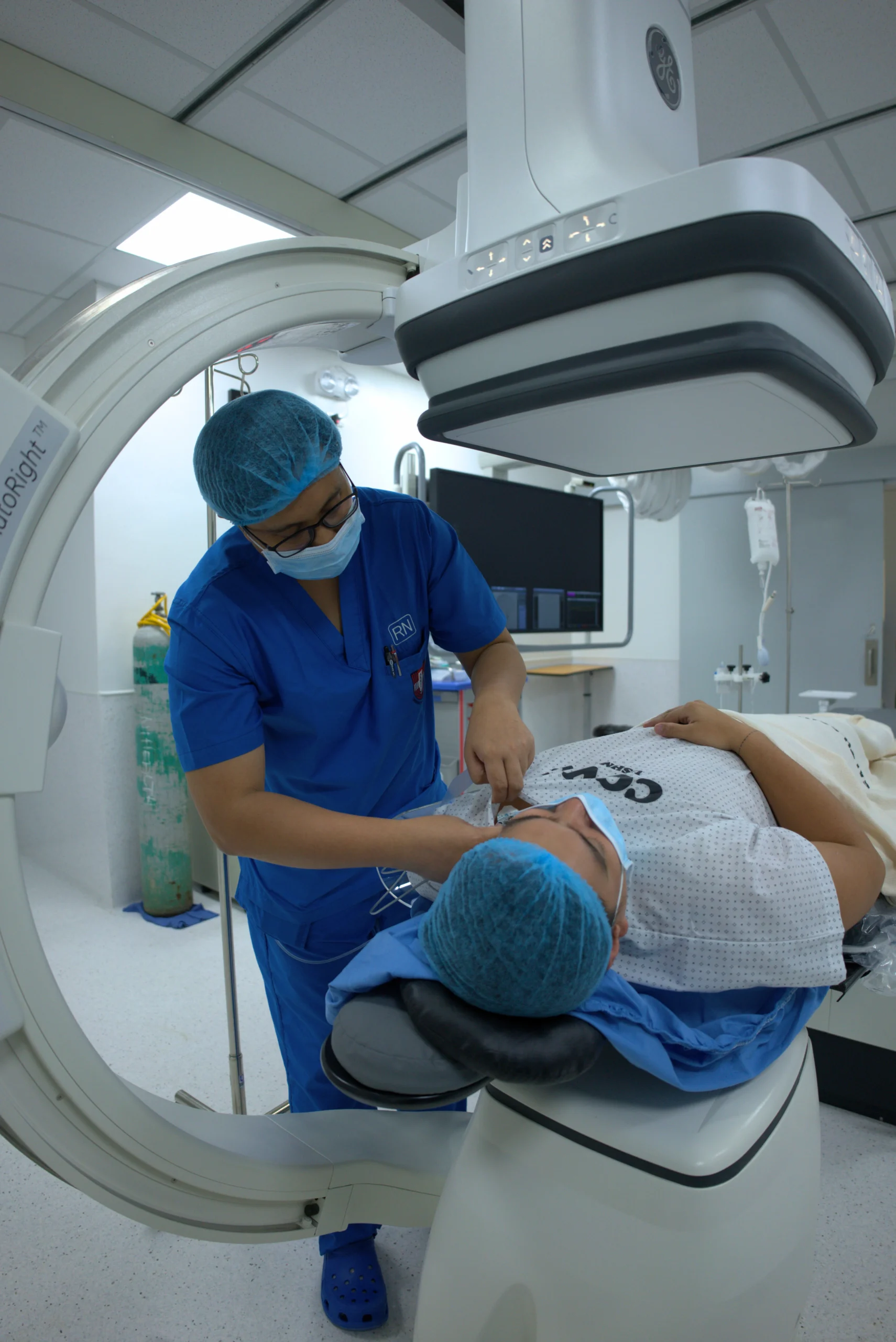
C-Arm
A C-Arm is a mobile, real-time X-ray imaging system named after its C-shaped arm that connects the X-ray source and the image detector.
Unlike normal X-rays that produce a single static image, the C-Arm lets our medical team view continuous, live X-ray images during a procedure, making it an essential tool for surgical guidance and interventional
procedures.
It is most commonly used during orthopedic surgeries (such as fracture repairs and joint replacements), pain management procedures, and minimally invasive surgeries where real-time imaging is very important for accuracy and patient safety.
The C-Arm allows our surgeons and specialists to make precise adjustments in real time, making
our procedures quicker and improving overall outcomes.
How to Prepare for X-ray, MRI, CT-Scan, or Ultrasound
The following preparation reminders for each procedure are general guidelines only. It is always best to ask your physician or the hospital staff to confirm the exact preparation requirements for your procedure.
X-ray Preparations
Remove all jewelry and metal such as necklaces, earrings, rings, zippers or belts. Having metal near the area to be scanned will block the X-rays and can make the image hazy, fuzzy, or cloudy.
MRI Preparations
Make sure to remove ALL metals in your body, including items like jewelry and hearing aids, before going into the room with the MRI machine. MRI machines have extremely strong magnets that can pull even big items such as hospital beds in. This is a crucial step to prevent any accidents or injuries from happening. It is also best to wear clothes that do not have any zippers, metal buttons, or belts.
Make sure to inform our staff of any metal medical implants such as cochlear implants, pacemakers, or aneurysm clips as most are not MRI-safe and are extremely risky to keep on your body.
Depending in the area/s of the body which will undergo the procedure.
For MRIs with Contrast, you will need to fast 4-6 hours before your procedure, similar to a CT-Scan.
It’s important to stay still as possible for 30-45 mins any movement will cause artifacts (blurry images). It will lead to repetition and make your total scan longer.
Ultrasound Preparations
For abdominal or pelvic ultrasound procedures, you may need to fast to stretch out your gallbladder or empty your stomach, or you may need to keep a full bladder to help move organs out of the way to create a clearer “imaging window”.
Other normal ultrasounds would typically not need any special preparations.
CT-Scan Preparations
General Preparations:
Like with normal X-rays procedures, you will need to remove all metal on your body such as jewelry and other accessories, to avoid affecting the images produced by the machine.
For With Contrast CT-Scan Procedures:
You will need to fast for 4-6 hours before your procedure. This is to prevent nausea or vomiting due to the contrast dye. If you are getting a CT-Scan in the abdominal area, this is to also make sure that your digestive tract has a clear view.
A laboratory test, specifically Creatinine, may be required depending on whether you have existing medical conditions that may be affected by the dye. This is usually ordered by your requesting doctor only when necessary.
Declare any allergies you may have, especially if you have an allergy to iodine, shellfish, or other previous contrast agents you may have encountered as the contrast dye contains iodine.
Why Choose Our Imaging Services?
State-of-the-art Equipment
We invest in the latest diagnostic imaging technology to meet industry standards and to maintain the quality of our imaging services. This includes a Digital X-ray machine, which emit 90% lower radiation than regular X-ray machines, a low-dose CT-Scan machine, and a high-resolution MRI machine. Our commitment to investing in modern equipment helps us ensure that the images we produce are clear, accurate, and precise so your doctor can give you the correct diagnosis and treatment while prioritizing your safety and comfort.
Board-Certified Radiologists and Technicians
Our licensed and board-certified radiologists and technicians review and interpret your results thoroughly and with high precision. They are highly trained to properly and safely operate our machines and equipment, while providing you with compassionate, patient-centered care during each procedure.
Timely and Accurate Results
Our streamlined workflow, interdepartmental coordination, and modern equipment allows us to capture clear images, store them securely, and interpret results more quickly. Faster turnaround times mean that your doctor can give you a proper diagnosis and treatment plan right away.
Board-Certified Radiologists and Technicians
From the moment you book your appointment until you leave our facility, your comfort and peace of mind are our priority. We focus on clear communication, fully explaining procedures and addressing concerns, especially regarding scans that may cause anxiety such as MRIs. We strive to provide our services with compassionate care and respect and make sure that your experience with us is as efficient, comfortable, and stress-free as possible.
Helpful Answers
Frequently Asked Questions
Do I need a doctor's request or referral for imaging tests?
Yes, you do. A doctor is needed because they evaluate your symptoms to ensure that the correct imaging test is done to protect patients from unnecessary radiation that may not be appropriate.The request guides the radiologist on what to look for so that the result can be accurate and useful for diagnosis.
Does my PhilHealth cover imaging tests?
Philhealth has benefit packages (case rates) for certain medical conditions which can include diagnostic test costs. However, it will not cover the total cost of your bill. For more information about your PhilHealth coverage, check our PhilHealth guide page.
Can I use my HMO health card to cover imaging tests?
Yes, you can. Present your HMO health card during billing to secure a Letter of Authorization (LOA) from your HMO for the specific procedure and contrast media before the exam can proceed. To learn more about how you can use your HMO at our hospital, you can view our HMO insurance guide page.
How long will it take to get my results?
Results for simpler exams such as X-rays and Ultrasounds can usually be released after only a few hours while results for more complex exams like CT-Scans or MRIs may take up to 3 days.
What do I need to prepare for an MRI or CT-Scan with contrast?
You will need to fast for 4-6 hours before your procedure and a blood test, specifically to check Creatinine, to make sure your kidneys can safely process the contrast dye.
What is the difference between an MRI and CT-Scan?
A CT-Scan uses X-rays and is better suited for imaging dense structures like bones, detecting acute injuries, or evaluating the chest and abdomen quickly.
An MRI uses magnets and radio waves (no radiation) and makes higher-resolution images of soft tissues like the brain, spinal cord, ligaments, and organs. Your doctor will determine which scan is most appropriate based on your symptoms and what needs to be evaluated.
How much does a Mammogram cost in the Philippines?
Mammogram prices vary by facility and whether it is a digital or analog mammogram. For our current Mammogram price, please contact our Information Department at +63 925 310 5666 or visit us at Unihealth Southwoods Hospital in Biñan, Laguna. We also offer Breast Ultrasound as an alternative or complementary screening tool.